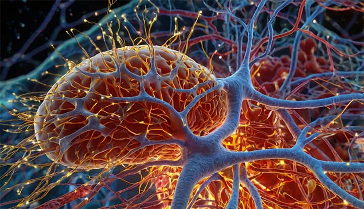
The autonomic nervous system is part of the nervous system responsible for controlling and regulating the body’s internal organs and systems, including the heart, breathing, digestion, and blood circulation, without conscious involvement. It is divided into the sympathetic and parasympathetic systems, which work together to maintain homeostasis and adapt the body to changes in the external and internal environments.
The importance of the autonomic nervous system in regulating vital processes cannot be overstated. It controls heart rate, blood pressure, breathing, digestion, excretions, body temperature, and many other functions. The ANS enables the body to adapt to changing conditions in the external and internal environments, coordinating the work of various organs and systems to maintain optimal functioning of the body as a whole.
Structure of the Autonomic Nervous System
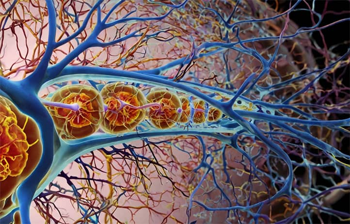
The autonomic nervous system is a complex network of neural pathways, which can be compared to a skilled conductor directing the orchestra of internal organs. Just as a conductor coordinates the sounds of different instruments, the ANS aligns the work of the heart, lungs, stomach, and other organs, creating a harmonious symphony of the body’s vital functions.
The evolution of the autonomic nervous system paralleled the increasing complexity of living organisms. From simple reflex arcs in primitive organisms to the most sophisticated regulatory mechanisms in mammals, this path reflects nature’s drive toward creating increasingly perfect adaptation systems.
Each part of the autonomic nervous system can be likened to a specialized department in a large corporation. The sympathetic division is the “rapid response” department, always ready to mobilize resources. The parasympathetic division is the “strategic planning” department, responsible for resource restoration and accumulation. And the metasympathetic division serves as “local management,” capable of making autonomous decisions at the level of individual organs.
Sympathetic Division
The sympathetic division of the ANS is often associated with the “fight or flight” response. It activates during stressful situations and prepares the body for intense physical or mental activity. The centers of the sympathetic nervous system are located in the thoracic and lumbar regions of the spinal cord.
Main functions of the sympathetic division include:
- increasing heart rate;
- raising blood pressure;
- dilating the bronchi;
- enhancing sweating;
- dilating the pupils;
- slowing intestinal peristalsis.
Parasympathetic Division
The parasympathetic division of the ANS, on the other hand, is responsible for the “rest and digest” state. It is active during calm conditions and contributes to the restoration of the body’s energy reserves. The centers of the parasympathetic nervous system are located in the brainstem and the sacral region of the spinal cord.
Main functions of the parasympathetic division include:
- reducing heart rate;
- lowering blood pressure;
- constricting the bronchi;
- enhancing the secretion of digestive glands;
- constricting the pupils;
- enhancing intestinal peristalsis.
Metasympathetic Division
The metasympathetic division of the ANS, also known as the enteric nervous system, is a network of neurons located in the walls of the gastrointestinal tract, urinary tract, and certain other internal organs. This division can function relatively autonomously, regulating the organs’ work even in the absence of signals from the central nervous system.
Main functions of the metasympathetic division include:
- regulating gastrointestinal motility;
- controlling the secretion of digestive glands;
- regulating local blood flow in organs.
Anatomy of the Autonomic Nervous System
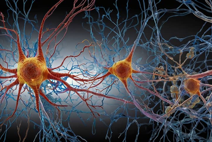
The anatomy of the autonomic nervous system is a fascinating world of neural connections and intersections. Like the root system of a mighty tree, autonomic nerve fibers permeate all organs and tissues, ensuring continuous connection with the central nervous system.
Ganglia in the autonomic nervous system can be compared to relay stations on the path of nerve impulses. These clusters of neurons not only transmit signals but also process information, making “local” decisions and adapting general commands to specific conditions in organs and tissues.
The history of studying the ANS anatomy is full of astonishing discoveries and unexpected findings. From the first descriptions of sympathetic trunks by Galen to modern neuroimaging methods, each stage of this journey has revealed new facets of the complexity and precision of autonomic regulation.
Central Divisions
The central divisions of the ANS are represented by structures located in the brain and spinal cord. These include:
- Hypothalamus: the highest center for autonomic function integration.
- Brainstem: contains centers of the parasympathetic nervous system.
- Spinal cord: contains centers of the sympathetic nervous system and some parasympathetic centers.
Peripheral Divisions
The peripheral divisions of the ANS include nerve fibers, ganglia, and nerve plexuses located outside the central nervous system.
Ganglia and Nerve Fibers
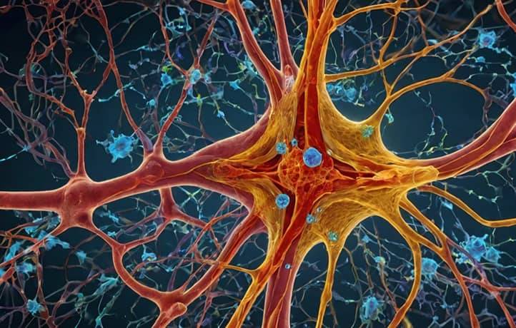
Ganglia are clusters of nerve cells that serve as relay stations for transmitting nerve impulses. In the autonomic nervous system, there are:
- Paravertebral ganglia: located along the spine and part of the sympathetic nervous system.
- Prevertebral ganglia: located in front of the spine and also part of the sympathetic system.
- Intramural ganglia: located in the walls of internal organs and part of the parasympathetic and metasympathetic systems.
The nerve fibers of the ANS are divided into preganglionic (before the ganglion) and postganglionic (after the ganglion). This structural feature is crucial for the autonomic nervous system’s functioning.
Physiology of the Autonomic Nervous System
The physiology of the autonomic nervous system involves a delicate structure of interactions and complex regulatory mechanisms. Like a conductor using baton movements to lead an orchestra, the ANS uses neurotransmitters to “conduct” the functions of internal organs.
Each neurotransmitter in the autonomic nervous system can be compared to a key that fits only a specific lock – a receptor. This specificity ensures accuracy and selectivity in autonomic regulation, allowing the body to finely tune its physiological processes.
Autonomic reflexes serve as the “autopilots” of our body. They help maintain homeostasis without conscious involvement, automatically responding to changes in the internal and external environments. These reflexes are the product of millions of years of evolution, refining mechanisms for rapid and effective adaptation to changing conditions.
Neurotransmitters and Receptors
In the ANS, the main neurotransmitters are acetylcholine and norepinephrine.
- Acetylcholine is released by all preganglionic fibers (both sympathetic and parasympathetic) and also by postganglionic parasympathetic fibers.
- Norepinephrine is released by most postganglionic sympathetic fibers.
These neurotransmitters interact with specific receptors on target cells, causing various physiological effects.
Signal Transmission Mechanisms
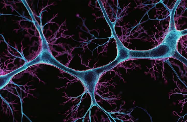
Signal transmission in the ANS follows this sequence:
- Generation of a nerve impulse in the central divisions of the ANS.
- Impulse conduction along the preganglionic fiber.
- Synaptic transmission in the ganglion.
- Impulse conduction along the postganglionic fiber.
- Neurotransmitter release in the synapse with the target cell.
- Interaction of the neurotransmitter with receptors and effect development.
Reflex Arc
Autonomic reflexes play a key role in regulating internal organs’ functions. The reflex arc of an autonomic reflex includes:
- Receptors that detect changes in the internal or external environment.
- Afferent (sensory) nerve fibers.
- Central division (in the spinal cord or brain).
- Efferent (motor) nerve fibers.
- Effector organ (smooth muscles, glands, etc.).
This system ensures a quick and appropriate response to various stimuli.
Functions of the Sympathetic Nervous System
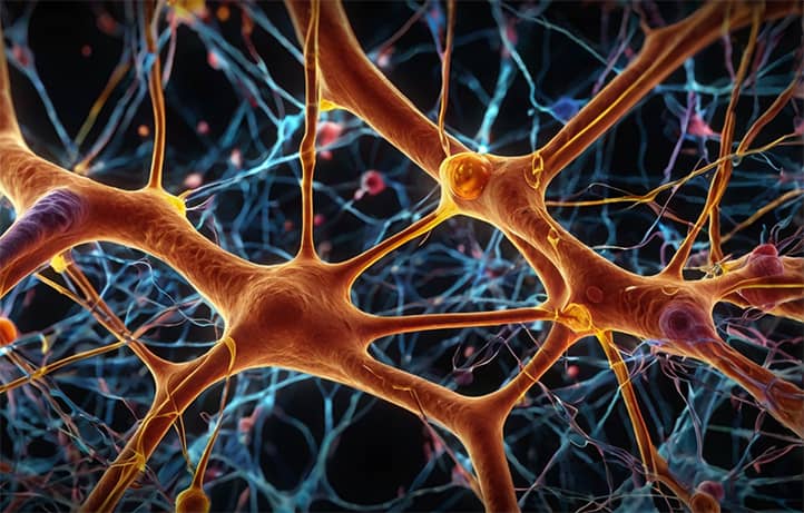
The sympathetic nervous system is the body’s “warrior,” always ready for battle. Just as a warrior musters all their strength before a fight, the sympathetic system activates all bodily resources in stressful situations.
Interestingly, the “fight or flight” response typical of sympathetic activation has deep evolutionary roots. This mechanism, developed by our distant ancestors to survive in a dangerous environment, has persisted into modern times. Today, it helps us cope not only with physical threats but also with psychological stressors.
The sympathetic nervous system’s effects on various organs can be compared to a coach motivating and directing a team to perform at its best. From an increased heartbeat to pupil dilation, all these effects aim to prepare the body for intense activity and overcoming challenges.
Fight or Flight Response
The “fight or flight” response is a complex physiological reaction of the body to perceived threats or stress. When the sympathetic nervous system is activated, it triggers:
- the release of adrenaline and noradrenaline from the adrenal glands;
- an increase in blood glucose levels;
- enhanced blood flow to muscles and the brain;
- suppression of digestive and excretory functions.
These changes prepare the body for intense physical or mental activity, increasing the chances of survival in critical situations.
Impact on Organs and Systems
The sympathetic nervous system has a diverse impact on various organs and systems:
- Cardiovascular system:
- increases heart rate and contraction strength;
- constricts blood vessels in most organs (except the heart and skeletal muscles);
- raises blood pressure.
- Respiratory system:
- dilates the bronchi;
- increases breathing rate.
- Digestive system:
- decreases stomach and intestine motility;
- reduces secretion of digestive glands;
- contracts sphincters.
- Urinary system:
- reduces urine formation;
- contracts the bladder sphincter.
- Endocrine system:
- stimulates glucose release from the liver;
- enhances secretion of adrenaline and noradrenaline.
- Skin:
- increases sweating;
- constricts blood vessels (paleness of the skin);
- causes piloerection (“goosebumps”).
- Eyes:
- dilates pupils;
- decreases accommodation.
These effects are aimed at mobilizing energy resources and preparing the body for active actions.
Functions of the Parasympathetic Nervous System
The parasympathetic nervous system is the body’s “peacekeeper.” Like a caring gardener, it creates optimal conditions for growth and recovery, ensuring efficient use of resources during rest.
The “rest and recovery” state typical of parasympathetic dominance can be compared to “recharging a battery.” In this mode, the body not only replenishes its energy but also performs “preventive maintenance” — regenerating tissues, synthesizing essential substances, and strengthening immunity.
It’s interesting to note that the parasympathetic system plays a key role not only in physiological processes but also in a person’s psycho-emotional state. Activation of the parasympathetic system contributes to reducing anxiety, improving mood, and enhancing cognitive functions, underscoring the close connection between “body” and “mind” in our organism.
Rest and Recovery State
In a calm state, parasympathetic nervous system activity dominates, promoting:
- reduced energy expenditure;
- restoration of energy reserves;
- enhanced anabolic processes;
- maintenance of homeostasis.
Impact on Organs and Systems
The parasympathetic nervous system has the following effects on organs and systems:
- Cardiovascular system:
- reduces heart rate and contraction strength;
- dilates blood vessels;
- lowers blood pressure.
- Respiratory system:
- constricts the bronchi;
- decreases breathing rate.
- Digestive system:
- increases stomach and intestine motility;
- boosts secretion of digestive glands;
- relaxes sphincters.
- Urinary system:
- increases urine formation;
- relaxes the bladder sphincter.
- Endocrine system:
- stimulates insulin production.
- Eyes:
- constricts pupils;
- enhances accommodation.
These effects aim to conserve and restore the body’s resources, as well as to support the normal functioning of internal organs during rest.
Interaction of the Sympathetic and Parasympathetic Systems
The interaction of the sympathetic and parasympathetic systems can be likened to a skillful dance where partners alternate or act together, creating a complex and harmonious pattern of movements. This “vegetative dance” finely tunes the body’s functions to meet current needs and environmental conditions.
Interestingly, the balance between sympathetic and parasympathetic activity isn’t static but constantly changes throughout the day. These fluctuations form unique “vegetative rhythms,” synchronized with circadian rhythms, which play a key role in regulating the sleep-wake cycle.
A disruption in the interaction between the sympathetic and parasympathetic systems can lead to various pathological conditions. For example, chronic stress can cause prolonged sympathetic dominance, which, in turn, may lead to hypertension, heart rhythm disorders, and other health problems.
Antagonism and Synergism
In most cases, the sympathetic and parasympathetic systems exert opposite (antagonistic) effects on organs. For example:
- On the heart: The sympathetic system increases heart rate, while the parasympathetic decreases it.
- On the bronchi: The sympathetic system dilates them, while the parasympathetic constricts them.
- On the digestive tract: The sympathetic system suppresses motility, while the parasympathetic enhances it.
However, in some cases, both systems display synergistic actions. For instance, they both participate in regulating sexual function, complementing each other at different stages of the sexual response.
Maintaining Homeostasis
The interaction between the sympathetic and parasympathetic systems plays a crucial role in maintaining homeostasis—the relative constancy of the body’s internal environment. This process includes:
- Continuous monitoring of internal environment parameters.
- Processing information in the central nervous system.
- Formulating responses via the sympathetic and parasympathetic systems.
- Adjusting organ and system functions to maintain optimal parameters.
The balance between sympathetic and parasympathetic activity constantly adjusts to meet the body’s needs and environmental conditions, ensuring appropriate adaptation to various situations.
The Enteric Nervous System
The enteric nervous system is like a “local manager” in the body, capable of making autonomous decisions at the level of individual organs. Just as local governments address city-level issues without always consulting the central government, the enteric system regulates organ functions independently of the central nervous system.
Evolutionarily, the enteric system is the oldest part of the autonomic nervous system. Its presence in primitive organisms lacking a developed central nervous system underscores the fundamental role of local regulation mechanisms in sustaining life.
Interestingly, the enteric system plays a key role in regulating the gastrointestinal (GI) tract—one of the body’s most complex and autonomous systems. The ability of the intestine to function even when completely disconnected from the central nervous system demonstrates the remarkable autonomy and “intelligence” of enteric regulation.
Characteristics and Functions
The primary characteristics of the enteric nervous system include:
- The ability to function autonomously, without direct control from the central nervous system.
- Possession of its own sensory, interneuron, and motor neurons.
- The ability to generate rhythmic activity.
The main functions of the enteric nervous system include:
- Regulating the motility of the gastrointestinal tract.
- Controlling the secretion of digestive glands.
- Regulating local blood flow in digestive organs.
- Coordinating the work of different parts of the digestive tract.
Role in Regulating GI Tract Function
The enteric nervous system plays a crucial role in ensuring the normal functioning of the gastrointestinal tract:
- Generates the basal electrical rhythm, determining the contraction frequency of GI tract smooth muscles.
- Coordinates peristaltic and segmental movements in the intestines.
- Regulates digestive juice secretion depending on food composition.
- Controls the absorption of nutrients.
- Facilitates local reflex responses to the stretching of organ walls or chemical stimuli.
Although the enteric system can function autonomously, it is also modulated by the sympathetic and parasympathetic systems, ensuring the integration of GI tract function with the body’s overall condition.
Autonomic Nervous System Disorders
Autonomic nervous system (ANS) disorders can be compared to malfunctions in the intricate mechanism of a clock. Just as the smallest glitch in one cogwheel can disrupt the entire timepiece, dysfunctions in autonomic regulation can cause imbalances throughout the body.
Autonomic disorders are often early indicators of various diseases, serving as the body’s “alarm signals.” The ability to recognize and accurately interpret these signals is a vital task of modern medicine, enabling the identification of pathological processes at early stages.
Treating autonomic disorders requires a comprehensive approach, taking into account the close connection between a person’s physical and psycho-emotional states. Methods for correcting autonomic disorders, from psychotherapy to neuromodulation, reflect a modern understanding of the body as a unified psychosomatic system.
Autonomic Vascular Dystonia (AVD)
Autonomic Vascular Dystonia (AVD) is a complex of functional disorders associated with the dysregulation of vascular tone. Key symptoms of AVD include:
- fluctuations in blood pressure;
- irregular heart rhythms (tachycardia, extrasystole);
- headaches;
- increased fatigue;
- disturbances in thermoregulation;
- emotional lability.
Causes of AVD vary and may include genetic predisposition, chronic stress, hormonal imbalances, and more.
Treating AVD typically involves a comprehensive approach:
- Lifestyle normalization (balancing work and rest, diet, physical activity).
- Psychotherapy.
- Medication (as necessary).
- Physiotherapy procedures.
Other Disorders
Beyond AVD, there are several other autonomic nervous system disorders:
- Postural Orthostatic Tachycardia Syndrome (POTS):
- characterized by a sharp increase in heart rate upon standing;
- may be accompanied by dizziness, weakness, fainting.
- Raynaud’s Syndrome:
- manifests as episodic spasms of blood vessels in the fingers or toes;
- causes blanching, then blueing and reddening of the fingers.
- Autonomic Neuropathy in Diabetes:
- leads to dysfunctions in cardiovascular, digestive, and urinary systems;
- can cause orthostatic hypotension and sweating disturbances.
- Horner’s Syndrome:
- characterized by ptosis (drooping of the upper eyelid), miosis (pupil constriction), and anhidrosis (absence of sweating) on the affected side of the face;
- arises from damage to sympathetic nerve fibers.
- Autonomic Crises (Panic Attacks):
- marked by acute episodes of autonomic dysfunction;
- accompanied by feelings of fear, anxiety, and autonomic symptoms (tachycardia, shortness of breath, sweating).
Diagnosing and treating these disorders require a comprehensive approach and often the involvement of specialists from different fields (neurologists, cardiologists, endocrinologists, etc.).
Conclusion
The autonomic nervous system represents an incredible example of biological regulatory perfection. Its ability to coordinate the functioning of numerous organs and systems, ensuring their harmonious operation in constantly changing external and internal environments, is truly remarkable. From maintaining internal balance to mobilizing resources in stressful situations, the autonomic nervous system plays a key role in sustaining life.
Researching the autonomic nervous system not only expands our understanding of human physiology but also opens up new possibilities for diagnosing and treating various diseases. Autonomic dysregulation underlies many pathological conditions, and correcting these disorders is often key to successful treatment.
The prospects for research into the autonomic nervous system are truly exciting. Exploring molecular mechanisms of autonomic regulation, developing new diagnostic and therapeutic methods for autonomic disorders, and studying the role of the ANS in aging processes – all these directions promise significant progress in medicine and physiology.
A deeper understanding of the link between the autonomic nervous system and psycho-emotional states opens new opportunities in psychosomatic medicine and stress management. Ultimately, studying the autonomic nervous system not only broadens our knowledge of bodily functioning but also brings us closer to unraveling one of nature’s greatest mysteries – the mechanisms that sustain life.
