In the modern world, mental disorders are becoming one of the most pressing public health issues. According to the World Health Organization (WHO), one in four people worldwide will experience some form of mental illness in their lifetime, such as depression, anxiety disorders, schizophrenia, or bipolar disorder.
Moreover, the prevalence of these conditions is increasing across all age groups, from adolescents to the elderly. The fast-paced lifestyle, constant stress, social isolation, environmental factors, and even the consequences of global crises are exacerbating the situation. Mental disorders not only reduce the quality of life for millions of people but also pose a significant economic burden on society, leading to loss of productivity and increased costs for treatment and rehabilitation.
In this context, early diagnosis and a precise understanding of the mechanisms underlying mental illnesses are of particular importance. Traditional diagnostic methods, based on clinical interviews and observations, often prove insufficiently accurate, especially in the early stages when symptoms are still mild. Furthermore, many mental disorders have similar manifestations, making it difficult to establish a correct diagnosis and choose effective treatment.
This is where modern technologies come into play, and among them, neuroimaging holds a special place. Neuroimaging enables scientists and doctors not only to detect changes associated with mental disorders but also to better understand their nature, opening new possibilities for developing personalized treatment approaches.
What is Neuroimaging?
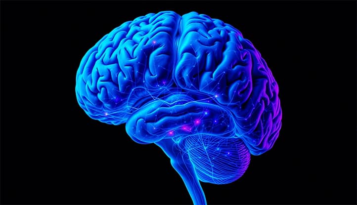
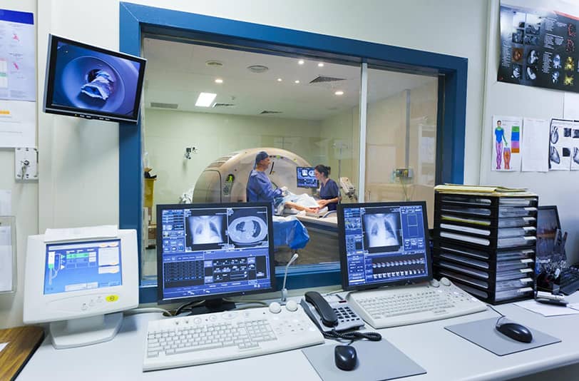
Neuroimaging is a set of methods that allow for the visualization of the structure, functions, and biochemical processes of the brain in real time. These technologies make it possible to “see” inside the brain without resorting to invasive procedures, making them indispensable in modern medicine and neuroscience.
The main neuroimaging methods include:
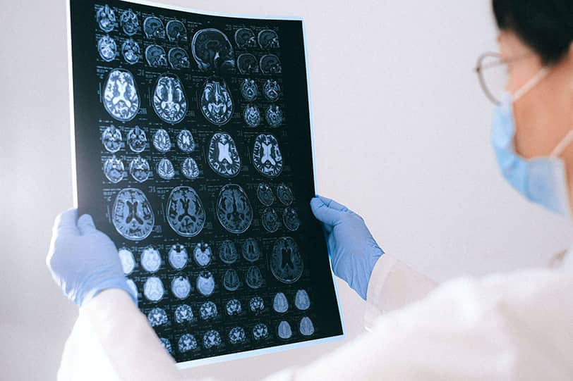
- Magnetic Resonance Imaging (MRI): Provides detailed, high-resolution images of the brain’s structure.
- Functional MRI (fMRI): Used to study the activity of different brain regions in response to stimuli or tasks.
- Positron Emission Tomography (PET): Helps investigate metabolic processes and the distribution of neurotransmitters, such as dopamine or serotonin, which is particularly important for understanding the mechanisms of mental disorders.
- Computed Tomography (CT): Less detailed than MRI but often used for emergency diagnostics, such as in cases of traumatic brain injuries.
- Electroencephalography (EEG): Records the brain’s electrical activity with high temporal precision, making it ideal for studying dynamic neural processes, such as sleep or epileptic seizures.
The role of neuroimaging in brain research cannot be overstated. It has been a true breakthrough in neuroscience. Thanks to neuroimaging, scientists have been able to identify brain regions responsible for various functions, such as speech, memory, emotions, and decision-making. For example, fMRI has revealed that the prefrontal cortex plays a key role in regulating emotions, while the hippocampus is crucial for memory formation.
Additionally, neuroimaging helps understand how the brain changes in various diseases. For instance, MRI can detect a reduction in hippocampal volume in Alzheimer’s disease, while changes in prefrontal cortex activity are observed in schizophrenia. These findings not only deepen our understanding of disease mechanisms but also open new avenues for developing treatments.
Neuroimaging is used for planning neurosurgical operations, monitoring the effectiveness of drug therapies, and even developing innovative approaches such as transcranial magnetic stimulation (TMS) or neurofeedback.
Thus, neuroimaging is not just a tool for obtaining brain images but a powerful method that allows researchers to explore brain function at all levels, from anatomical to molecular. It serves as a bridge between fundamental science and clinical practice, helping not only to better understand how the brain is structured and functions but also how to treat it.
Neuroimaging as a Diagnostic Tool
Modern neuroimaging techniques not only allow us to visualize the anatomical features of the brain but also assess its functional activity, blood flow, metabolism, and even neurochemical processes. This makes neuroimaging a universal tool used for both diagnosis and treatment monitoring.
For example, neuroimaging can detect early signs of neurodegenerative diseases long before clinical symptoms appear. Additionally, it helps differentiate between mental disorders, which is particularly important for selecting the right treatment strategy.
How Does Neuroimaging Work?
Neuroimaging is based on various physical and biological principles that enable the creation of highly accurate brain images. For instance:
- Magnetic Resonance Imaging (MRI): Uses powerful magnetic fields and radiofrequency pulses to create detailed images of the brain’s structure. This method relies on the phenomenon of nuclear magnetic resonance, where hydrogen atoms in brain tissues respond to the magnetic field, generating signals that are then converted into images.
- Positron Emission Tomography (PET): Utilizes radioactive tracers injected into the body, which accumulate in active brain regions, allowing visualization of metabolic processes.
- Computed Tomography (CT): Operates using X-rays that pass through brain tissues, creating cross-sectional images.
- Electroencephalography (EEG): Records the brain’s electrical activity via electrodes placed on the scalp, enabling the study of neural processes in real time.
The Difference Between Structural and Functional Imaging
Structural imaging, such as MRI and CT, focuses on studying the brain’s anatomy. It helps identify changes in tissue volume, shape, and density, which is particularly important for diagnosing tumors, injuries, and neurodegenerative diseases. For example, structural MRI can detect hippocampal atrophy, a hallmark of Alzheimer’s disease.
Functional imaging, such as fMRI and PET, concentrates on brain activity. It shows which brain regions are activated during specific tasks or at rest. For instance, fMRI is used to study prefrontal cortex dysfunction in depression or schizophrenia. Thus, structural and functional imaging complement each other, providing a comprehensive picture of the brain’s condition.
Examples of Use in Diagnosis
Neuroimaging plays a key role in diagnosing mental disorders by identifying specific changes in the brain. For example:
- In depression, fMRI can reveal reduced activity in the prefrontal cortex and hyperactivity in the amygdala, associated with impaired emotion regulation.
- In schizophrenia, structural MRI often shows a reduction in gray matter volume in the temporal and frontal lobes, while PET reveals imbalances in the dopamine system.
- Bipolar disorder also has its neuroimaging markers, such as changes in amygdala volume and disrupted connectivity between different brain regions.
These findings help not only refine diagnoses but also develop personalized treatment approaches.
Early Markers of Neurodegenerative Diseases
Neuroimaging is particularly important for the early diagnosis of neurodegenerative diseases such as Parkinson’s, Alzheimer’s, and others. For example:
- MRI can detect a reduction in hippocampal volume, one of the earliest signs of Alzheimer’s disease.
- PET with specialized radioactive tracers can identify the accumulation of amyloid plaques and tau proteins, which are considered key biomarkers of Alzheimer’s.
Early diagnosis through neuroimaging allows treatment to begin at a stage when symptoms have not yet manifested, significantly improving prognosis and patients’ quality of life.
Neuroimaging and Understanding the Mechanisms of Mental Disorders
Mental disorders such as depression, schizophrenia, anxiety, and bipolar disorder have long remained a mystery to scientists and doctors. Their symptoms are often difficult to interpret, and the mechanisms underlying them have remained hidden from researchers. However, with the advent of neuroimaging, the situation has changed dramatically.
This method has allowed us to look inside the living brain and see how it functions in both normal and pathological states. Neuroimaging has become a key tool for studying brain activity, its structure, and neurochemical processes, significantly deepening our understanding of mental disorders.
Studying Brain Activity and Dysfunctions in Neural Networks
Neural networks are complex systems of interaction between different brain regions responsible for cognitive, emotional, and behavioral functions. In mental disorders, these networks often malfunction, leading to impairments in thinking, emotions, and behavior.
Neuroimaging, particularly fMRI, allows researchers to study the activity of neural networks in real time. For example, in depression, reduced activity is observed in the default mode network, which is responsible for self-reflection and processing internal experiences. At the same time, anxiety disorders often show hyperactivity in the network associated with processing fear and threats. These findings help scientists understand which specific networks are disrupted in a given disorder and develop methods for their correction.
Hyperactivity of the Amygdala in Anxiety Disorders
The amygdala is a small brain region that plays a key role in processing emotions, especially fear and anxiety. In anxiety disorders, such as generalized anxiety disorder or panic attacks, the amygdala often becomes hyperactive. This can be observed using fMRI, which shows increased activity in this region in response to neutral or even positive stimuli.
Such hyperactivity leads individuals to perceive ordinary situations as threatening, triggering anxiety symptoms. Understanding this mechanism allows for the development of treatments aimed at reducing amygdala activity, such as cognitive-behavioral therapy or medications.
Hippocampal Volume Changes in Depression
The hippocampus is a brain region crucial for memory formation and emotion regulation. In depression, a reduction in hippocampal volume is often observed, linked to chronic stress and elevated cortisol levels. These changes can be detected using structural MRI.
A reduction in hippocampal volume not only impairs memory but also complicates emotion regulation, exacerbating depressive symptoms. Understanding this connection helps in developing treatments aimed at protecting and restoring the hippocampus, such as antidepressants or physical activity.
Prefrontal Cortex Dysfunction in Schizophrenia
The prefrontal cortex is a brain region responsible for higher cognitive functions, such as planning, decision-making, and impulse control. In schizophrenia, disruptions in the structure and function of the prefrontal cortex are often observed. For example, structural MRI can reveal a reduction in gray matter volume in this region, while fMRI shows decreased activity during cognitive tasks.
These dysfunctions lead to symptoms of schizophrenia, such as impaired thinking, reduced motivation, and social isolation. Understanding these changes helps in developing treatments aimed at improving prefrontal cortex function, such as cognitive rehabilitation or medications.
Neurochemical Processes: Using PET to Study Dopamine and Serotonin Systems
Dopamine and serotonin are neurotransmitters that play a key role in regulating mood, motivation, and behavior. In mental disorders such as depression, schizophrenia, and bipolar disorder, dysfunctions in these systems are often observed. PET allows researchers to study the distribution and activity of dopamine and serotonin receptors in the brain.
For example, in schizophrenia, hyperactivity of the dopamine system in the striatum is often observed, associated with symptoms such as hallucinations and delusions. In depression, on the other hand, reduced activity of the serotonin system is common, leading to mood disturbances and apathy. These findings help in developing medications that restore neurotransmitter balance, such as antipsychotics or antidepressants.
Neuroimaging in the Development of Treatment Methods
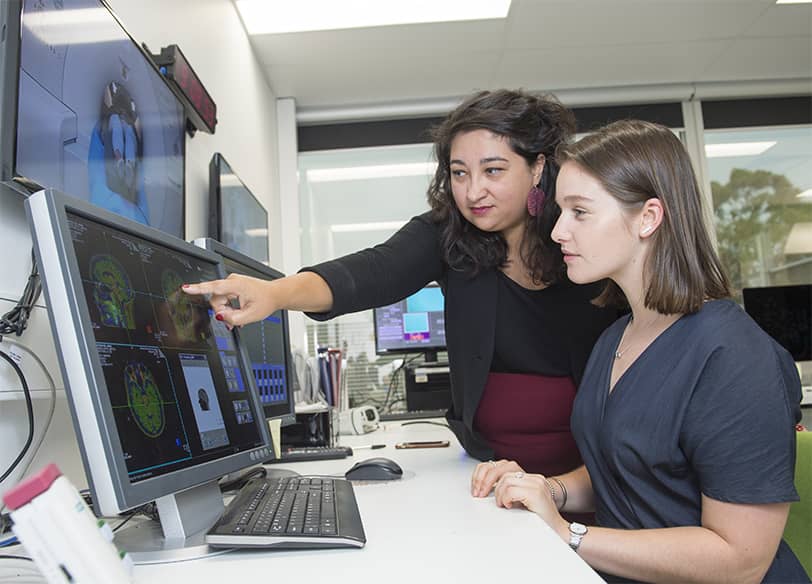
Modern medicine is increasingly moving towards personalized approaches, where treatment is tailored not only based on diagnosis but also considering the individual characteristics of the patient. Neuroimaging plays a key role in this process by providing unique data on the structure, function, and biochemistry of the brain.
This data allows doctors not only to better understand the nature of the disease but also to develop individualized treatment plans that take into account the specific brain functioning of each patient. Moreover, neuroimaging is used to monitor the effectiveness of therapy, enabling the assessment of changes in the brain after treatment and adjusting it as necessary.
Personalized Medicine
Personalized medicine is an approach where treatment is tailored based on the individual characteristics of the patient, including genetics, biochemistry, and the functional state of the brain. Neuroimaging plays a crucial role in this process by providing data on how the brain of a specific individual functions.
For example, fMRI can identify which brain regions are underactive in depression or overactive in anxiety disorders. This data allows doctors to select medications that target these specific areas or recommend therapies aimed at correcting the identified dysfunctions.
Additionally, neuroimaging helps differentiate patients with similar symptoms but different disease mechanisms. For instance, in depression, some patients may exhibit reduced activity in the prefrontal cortex, while others may show hyperactivity in the amygdala. In the first case, antidepressants that stimulate prefrontal cortex activity may be effective, while in the second case, medications that reduce anxiety may be more appropriate. Thus, neuroimaging not only improves treatment effectiveness but also helps avoid unnecessary side effects.
Monitoring Treatment Effectiveness
One of the key challenges in treating mental disorders is assessing the effectiveness of therapy. Neuroimaging provides objective data that allows evaluation of how treatment affects the brain. For example, fMRI can show changes in prefrontal cortex activity after a course of antidepressants or reduced hyperactivity in the amygdala following treatment for anxiety disorders.
Furthermore, neuroimaging helps identify patients who do not respond to standard treatment. For instance, some patients with depression may not show improvement in prefrontal cortex activity after taking antidepressants. In such cases, doctors can adjust the treatment, for example, by adding psychotherapy or switching to a different medication.
New Approaches to Treatment
Neuroimaging opens up new possibilities for developing innovative treatment methods, such as transcranial magnetic stimulation (TMS) and neurofeedback. TMS is a method that uses magnetic pulses to stimulate specific brain regions. Neuroimaging, particularly fMRI, is used to precisely identify these regions.
For example, in depression, TMS is often targeted at stimulating the dorsolateral prefrontal cortex, which may be underactive. Neuroimaging data allows for precise localization of the stimulation site and customization of the procedure parameters for each patient, making the treatment more effective.
Neurofeedback is a method where patients learn to regulate their brain activity using real-time feedback. With EEG, patients can see how their brain activity changes and learn to control it.
Limitations and Ethical Issues
Despite its immense potential, neuroimaging faces a number of limitations that can hinder its widespread application in clinical practice and scientific research. These limitations are both technical and ethical in nature.
Technical and Methodological Limitations
One of the main challenges of neuroimaging is the complexity of interpreting the data. The brain is an extremely complex organ, and its functioning depends on numerous factors, such as genetics, environment, the patient’s current state, and even the time of day.
For example, the activity of a specific brain region identified through fMRI may be associated with multiple processes, and it is not always clear how exactly it relates to the symptoms of a disorder. Additionally, neuroimaging data often require complex statistical processing, which can lead to errors or false conclusions. These difficulties make data interpretation a challenging task that requires high expertise and caution.
High Costs and Limited Accessibility
Neuroimaging is an expensive method that requires sophisticated equipment and highly qualified personnel. For instance, the cost of an MRI machine can reach several million dollars, and its maintenance and operation also entail significant expenses. This makes neuroimaging inaccessible to many medical institutions, particularly in developing countries or regions with limited resources.
Moreover, even in developed countries, access to neuroimaging may be limited due to long waiting lists and high procedure costs. This creates inequality in access to modern diagnostic and treatment methods, which is particularly critical for patients with mental disorders, who often already face social isolation and a lack of resources.
Data Privacy Concerns
Neuroimaging generates vast amounts of data that may contain personal information about the patient, such as anatomical features, functional characteristics of the brain, and even potential disease risks. This raises important questions about data confidentiality and protection.
For example, if neuroimaging data falls into the hands of insurance companies or employers, it could lead to discrimination against patients. Additionally, the use of neuroimaging data in scientific research requires clear patient consent and adherence to strict ethical standards. However, even with consent, questions remain about how this data will be stored, used, and protected in the long term.
Risks of Overdiagnosis and Stigmatization
Neuroimaging can detect changes in the brain that are not always linked to clinically significant symptoms. This creates a risk of overdiagnosis, where patients are diagnosed based on neuroimaging data even in the absence of clear symptoms of a disorder.
For instance, the detection of amyloid plaques in the brain may indicate a risk of Alzheimer’s disease, but it does not always mean that the person will develop the condition. Such overdiagnosis can cause unnecessary anxiety and stress in patients.
The Future of Neuroimaging in Psychiatry
Neuroimaging already plays a key role in the study and treatment of mental disorders, but its potential is far from exhausted. With the development of new technologies and data analysis methods, it promises to become an even more powerful tool that could revolutionize psychiatry.
In the coming years, we can expect significant progress in the fields of artificial intelligence (AI) and machine learning (ML), which will enable the analysis of neuroimaging data with unprecedented accuracy and speed. Additionally, the emergence of new imaging methods with higher resolution and sensitivity will open up new possibilities for studying the brain at the molecular and cellular levels.
These advancements will not only improve our understanding of mental disorders but also make neuroimaging accessible to a broader range of patients, integrating it into everyday clinical practice.
Technological Advancements
Artificial intelligence (AI) and machine learning (ML) are beginning to play an important role in the analysis of neuroimaging data. These technologies allow for the processing of vast amounts of information, identifying complex patterns that may be invisible to the human eye.
For example, ML algorithms can analyze fMRI data to detect early signs of mental disorders, such as changes in neural network activity. This is particularly important for diseases that are difficult to diagnose in their early stages, such as schizophrenia or bipolar disorder.
Moreover, AI can assist in personalizing treatment by analyzing neuroimaging data and predicting which therapeutic methods will be most effective for a specific patient.
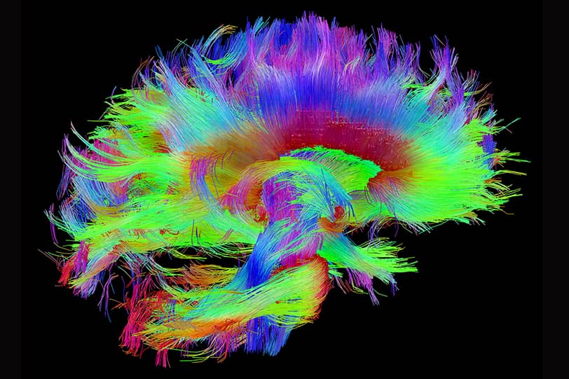
Modern neuroimaging methods, such as MRI and PET, already offer high precision, but their capabilities continue to expand. New methods are being developed that allow for imaging the brain with even higher resolution, which is especially important for studying small structures like the hippocampus or amygdala.
Technologies are also emerging that enable the study of the brain at the molecular level, such as new radioactive tracers for PET or nanotechnology-based methods. These advancements will help scientists better understand the mechanisms of mental disorders and develop more precise diagnostic and treatment methods.
Prospects for Patients
One of the most promising prospects for neuroimaging is the potential for early diagnosis and prevention of mental disorders. Already, neuroimaging can detect changes in the brain that precede the onset of clinical symptoms.
In the future, this could lead to the creation of screening programs that identify at-risk groups and initiate treatment at the earliest stages, when it is most effective. Additionally, neuroimaging could be used to monitor the effectiveness of preventive measures, such as cognitive training or lifestyle changes.
Integration of Neuroimaging into Everyday Clinical Practice
Currently, neuroimaging is primarily used in research and specialized clinics, but in the future, it could become part of everyday clinical practice. The reduction in equipment costs and the development of portable devices, such as simplified versions of EEG or fMRI, will make neuroimaging more accessible to a wider range of patients.
This will allow doctors to make more informed decisions based on objective data about a patient’s brain state. For example, neuroimaging could be used to tailor individual medication dosages or to assess the effectiveness of therapy in real time. Furthermore, integrating neuroimaging with other diagnostic methods, such as genetic testing, will enable the development of comprehensive approaches to treating mental disorders.
Conclusion
Neuroimaging has already proven its value as a powerful tool for understanding and treating mental disorders. Today, it allows us to look inside the living brain, observe its structure, activity, and biochemical processes, opening new horizons in the diagnosis, treatment, and prevention of mental illnesses.
Thanks to neuroimaging, we can not only detect changes associated with disorders such as depression, schizophrenia, or anxiety disorders but also better understand their nature, making treatment more precise and personalized. However, despite all the achievements, the potential of neuroimaging is far from fully realized.
Modern technologies, such as artificial intelligence and machine learning, promise even greater accuracy and detail. These advancements pave the way for early diagnosis, where treatment can begin before serious symptoms appear, and for more effective therapeutic methods that take into account the individual characteristics of each patient.
Nevertheless, to fully realize the potential of neuroimaging, further research and active integration of these technologies into clinical practice are needed. This requires not only financial investment but also the training of specialists capable of working with modern equipment and interpreting complex data. Additionally, it is crucial to address ethical issues related to data privacy and the risks of overdiagnosis to ensure that the use of neuroimaging is not only effective but also safe for patients.
The integration of neuroimaging into everyday medical practice could be a significant step toward more personalized and precise medicine, where treatment is based not only on symptoms but also on a deep understanding of disease mechanisms. This is especially important for psychiatry, where many disorders remain a mystery, and treatment methods are often insufficiently effective.
Thus, neuroimaging is not just a technology but a key to a new understanding of mental health and illness. It opens up the possibility of not only better treatment but also the prevention of mental disorders, improving the quality of life for millions of people worldwide.